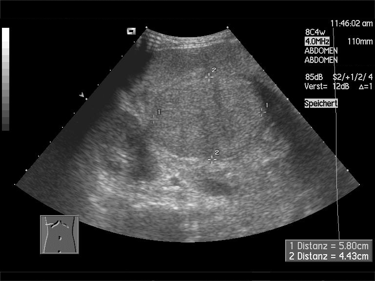
liver cirrhosis. liver cirrhosis. July 19, 2010
![[RU] Ultrasound image description. Liver cirrhosis. [RU] Ultrasound image description. Liver cirrhosis.](http://www.medison.ru/uzi/img/p97.jpg)
[RU] Ultrasound image description. Liver cirrhosis.
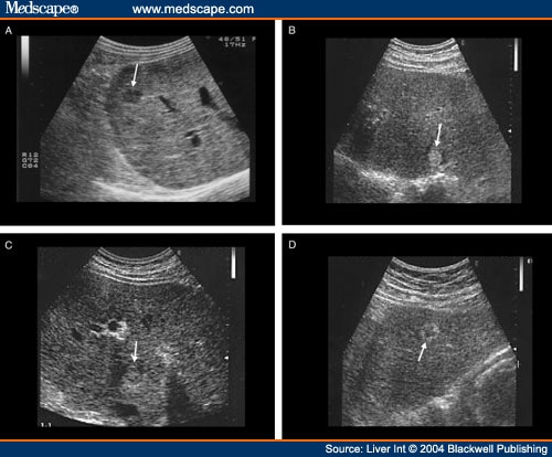
carcinomas detected in patients with liver cirrhosis: hypoechoic (A),

Diagnosis for Liver Cirrhosis by Ultrasound Images
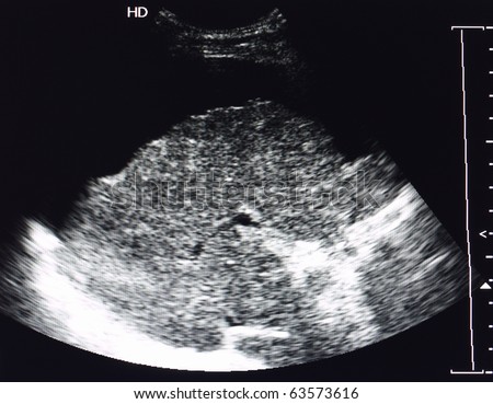
stock photo : ultrasound image of liver cirrhosis and ascites

Although most commonly due to cirrhosis and severe liver disease,

Sonograms are used to diagnose and monitor fatty liver disease. Ultrasound
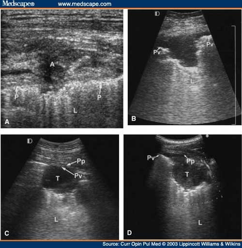
(A) Ultrasound (US) shows a chest wall abscess in a patient with liver

Typical human liver image. Click for larger image. Standard ultrasound

Cirrhosis of liver with portal vein thrombosis:

9) Cirrhosis of liver and hepatorenal syndrome in neonate:

The loss of normal liver tissue slows the processing of nutrients, hormones,

A chronic liver disease which causes damage to liver tissue,
TREATMENT OF CIRRHOSIS OF THE LIVER. Treatment is symptomatic and usually
Cirrhosis of the Liver - MedPix™: 19181 - Medical Image Database and Atlas

These findings were compatible with liver cirrhosis (Fig. 2).

This is the most accurate way to diagnose cirrhosis. In a liver biopsy,
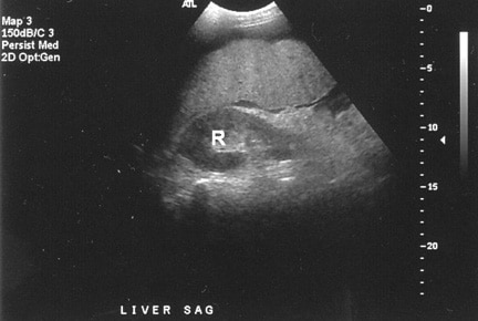
Advanced cirrhosis. A nodular liver, echogenic in comparison to renal

A cirrhosis liver has coarser echo-texture in (a) the liver than that in (b)

liver- What is liver cirrhosis


